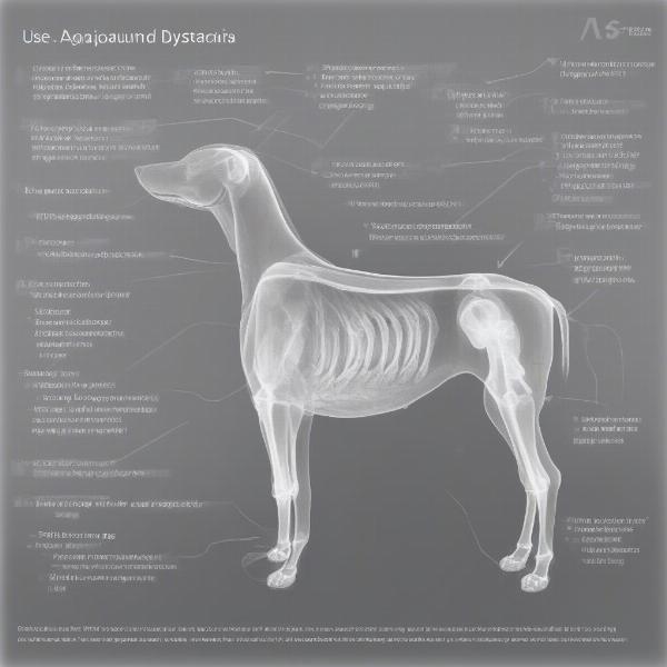
Hip dysplasia is a common skeletal condition affecting dogs, particularly larger breeds. It’s a developmental disease where the hip joint doesn’t form correctly, leading to instability and potentially debilitating arthritis. A crucial tool in diagnosing hip dysplasia is the use of x-rays. Understanding the process and what these x-rays reveal can help owners navigate this challenging condition and ensure the best possible care for their canine companions.
What to Expect During Hip Dysplasia X-Rays
The process of taking hip dysplasia x-rays involves precise positioning to get clear images of the hip joints. Your veterinarian will likely sedate or anesthetize your dog to ensure they remain still and relaxed during the procedure. This not only provides higher quality x-rays but also minimizes stress for your dog. The x-rays themselves are quick, and your dog will be monitored throughout the process. Your vet will then analyze these images to assess the degree of hip dysplasia present.
Decoding the X-Ray: What the Images Reveal
Hip dysplasia x-rays allow veterinarians to evaluate the structure and alignment of the hip joint. Key features they look for include the shape of the femoral head (the ball of the hip joint), the depth of the acetabulum (the socket of the hip joint), and the presence of any signs of osteoarthritis, like bone spurs or joint space narrowing. The severity of hip dysplasia is often graded based on these findings, which helps determine the appropriate treatment plan.
 Hip Dysplasia X-Ray Interpretation
Different X-Ray Techniques for Hip Dysplasia Diagnosis
There are several x-ray techniques employed in diagnosing hip dysplasia. The standard view is the ventrodorsal extended leg view, which provides a comprehensive overview of the hip joint. Other views, like the PennHIP method, which assesses joint laxity, might also be used for a more detailed analysis, especially in younger dogs. Your veterinarian will choose the most appropriate method based on your dog’s age, breed, and suspected severity of the condition.
Why are hip dysplasia x-rays important?
Hip dysplasia x-rays are crucial for accurate diagnosis and treatment planning. They allow veterinarians to visualize the joint structure and assess the severity of the condition. This information is essential for determining the best course of action, whether it’s medical management, surgery, or a combination of both. Early diagnosis through x-rays can significantly improve a dog’s long-term prognosis.
What is the best age for hip dysplasia x-rays?
While hip dysplasia can be suspected based on clinical signs, definitive diagnosis usually requires x-rays. For most breeds, x-rays are recommended at two years of age. However, some breeds prone to hip dysplasia might benefit from earlier screening. Discuss with your veterinarian the optimal timing for x-rays based on your dog’s breed and any observed symptoms.
Managing Hip Dysplasia: Beyond the X-Ray
While x-rays are essential for diagnosis, managing hip dysplasia is a multifaceted approach. Treatment options range from pain management and physical therapy to surgical interventions. Maintaining a healthy weight, providing regular low-impact exercise, and using joint supplements can also play a significant role in improving your dog’s quality of life. muscle deterioration in dogs and old dog weak back legs are related issues that should be addressed.
Conclusion: Hip Dysplasia X-Rays – A Vital Step
Hip dysplasia x-rays are a vital tool in diagnosing and managing this common canine condition. By understanding the process and what the images reveal, owners can make informed decisions about their dog’s care. Early diagnosis through hip dysplasia x-rays, combined with appropriate treatment and management strategies, can significantly improve a dog’s comfort and mobility, allowing them to live a full and active life. If you suspect your dog may have hip dysplasia, consult your veterinarian to discuss x-ray screening and the best course of action. bone disease in dogs can be a contributing factor and should be considered.
FAQs
-
Do hip dysplasia x-rays hurt my dog? No, the x-ray itself is painless. However, sedation or anesthesia is usually required to ensure proper positioning, which carries minimal risks.
-
How much do hip dysplasia x-rays cost? The cost varies depending on your location and veterinary clinic. It’s best to contact your vet for a specific quote.
-
Can hip dysplasia be cured? While there’s no cure, various treatments can manage the condition and improve your dog’s quality of life. soft tissue damage dog can be a related concern.
-
Are certain breeds more prone to hip dysplasia? Yes, larger breeds like German Shepherds, Golden Retrievers, and Labrador Retrievers are more susceptible. bernese mountain dog rescue uk is a good resource if you are interested in this breed.
-
What are the signs of hip dysplasia in dogs? Common signs include lameness, difficulty rising, stiffness, and decreased activity.
ILM Dog is a leading online resource dedicated to providing dog owners with expert advice and information on all aspects of canine care, from breed selection and health to training and nutrition. We offer a wealth of resources to help you navigate every stage of your dog’s life, ensuring their health, happiness, and well-being. From understanding crucial diagnostics like hip dysplasia x-rays to selecting the right products and accessories, ILM Dog is your trusted companion on this journey. Contact us at [email protected] or +44 20-3965-8624 for personalized guidance.